Objective
Spatial resolution in a radiograph, just as in a television, lets the viewer see small and narrow objects. For instance, an endo file or a PDL space can be too narrow for a lower resolution sensor to image. However, a higher resolution sensor has smaller and more numerous pixels, generating images which distinguish small features from their surroundings. Also, since trabecular bone is made up of many fine structures, superior spatial resolution provides more information about bone health.
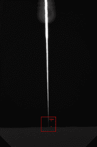
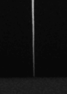
An easy to obtain measurement is whether the sensor can image the tip of the #6 endo file. This file’s tip is approximately 60 microns in diameter, far smaller than lesions and typical anatomic structures. Since this is the smallest, narrowest object the clinician is interested in, this measurement is a good standard for spatial resolution.
Design
XDR’s pixel size is 19 microns. This provides a theoretical spatial resolution of 26.3 line pairs/mm. However, no sensor ever achieves its theoretical spatial resolution. In tests under optimal conditions, the Anatomic Sensor achieves 20 line pairs/mm. (Figure 2 demonstrates that one can see, on good monitors, the grating’s 5 dark lines as distinct all the way down to the 20-line-pair indicator.)
 |
 |
Results
In a peer reviewed study, using more realistic conditions, the Anatomic Sensor’s spatial resolution is in the top tier. In Figures 3 and 4, one can locate the tip of the #6 file because it is against a piece of acrylic, thus demonstrating that the sensor successfully images that very narrow tip.
 |
 |
Conclusion
The Anatomic Sensor achieves a practical standard of spatial resolution in dental radiography: being able to see the tip of the a #6 file. With that, the ability to see anatomic structures is enhanced, improving diagnosis.
|
|
|

