Objective
Soft tissue often shows up poorly in both traditional and digital radiographs. The problem is accentuated by the fact that a digital radiograph on a computer monitors has less dynamic range (approximately 1000:1) than does a film in a light box (approximately 10,000:1). Spot filters are sometimes used to show soft tissue, but these filters often obscure contrast in regions outside the density range of interest. A filter is needed which can accentuate the contrast in regions of low radiographic density, while still maintaining visualization of the denser regions in the radiograph.
Design
Using proprietary adaptive dynamic range compression methods similar to those in the Caries/Endo filter, XDR designed the Perio filter. This filter improves visualization of the soft tissue while avoiding the drawbacks of spot filters. The Perio filter thus enables the clinician to see all anatomic regions of the radiograph, providing the contextual information needed to make a more complete radiographic diagnosis.
Results
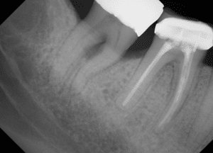
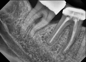
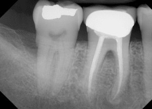
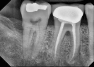
The figures illustrate how the low density interradicular regions of the radiograph are shown more completely in the Perio Filtered image. Note the greater radiographic height of the crestal bone and appearance of the gingival tissue in the filtered image.
|
|
|
|
|
|
Conclusions
Although all final diagnoses should be performed with un-filtered images, tools such as the Perio filter help the diagnostician see radiographic features they might otherwise miss. And, unlike some dental imaging programs, which concentrate on contrast in high density (hard tissue) regions at the expense of low density (soft tissue) regions, XDR’s Perio filter helps to elucidate the anatomic boundaries of the soft tissue regions in a radiograph, while preserving contextual information in the rest of the radiograph. This provides for a more complete clinical conclusion.


